What Is Veterinary Diagnostic Imaging For Felines?
Veterinary diagnostic imaging includes radiographs (x-rays), ultrasound, MRIs and CT scans, all of which are used as diagnostic tools to collect information on your cat's health. The vast majority of imaging is non-invasive and completely painless. However, some imaging may require sedation or anesthesia because the cat must be kept still to allow for adequate images to be produced. Veterinarians use these images to collect information on your cat to help them to make a medical and sometimes surgical plan.
When Is Diagnostic Imaging Necessary For Your Cat?
 After your veterinarian has examined your cat, he or she may want to begin to collect more information that will lead to a diagnosis and then, a treatment plan. X-rays are usually the first line of imaging. The x-ray may lead to a diagnosis that allows them to move forward with a plan. However, sometimes the next step may be ultrasound to get a more thorough or specific look at a particular area of the body.
For instance, if your cat is vomiting and feeling ill, your veterinarian may take an x-ray to look for possible causes such as obstruction of intestines or an obvious foreign body. The x-ray may show some signs suggestive of intestinal obstruction, however, before proceeding to surgery, it would be prudent in some cases to follow with an abdominal ultrasound. The ultrasound can give more detail of the area and therefore allow more confidence in the treatment plan to move forward with surgery. Sometimes, x-rays and ultrasound allow for a definitive diagnosis but other times they will simply add more information to help put the puzzle together for the best treatment plan for your cat.
The two types of diagnostic imaging used in our hospital to assist in diagnosis are radiographs and ultrasound. If the case requires the need for advanced imaging like CT or MRI, we will discuss referral to a local specialty hospital for this.
More information on each of these types of imaging is provided below.
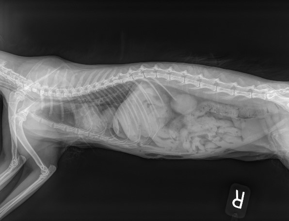
Radiographs
X-rays have been in use throughout the medical community for many decades. Radiogrpahs are by far the most regularly used form of diagnostic imaging in the veterinary industry because they are cost-effective (comparatively speaking), and they can accurately diagnose the state of skeletal structure and composition, large body cavities, and the presence of many foreign objects. X-rays are totally painless, but some cats can benefit from sedation to reduce anxiety and stress with positioning.
Radiographs usually proceed as follows:
- The cat is placed on the x-ray table
- A technician positions the x-ray machine so that the x-ray beam targets only the area of interest
- Modern x-ray equipment allow for low levels of radiation and when used only occasionally are perfectly safe for your cat
- Because x-rays are static images, the procedure usually requires less time than a procedure like an MRI
X-rays have traditionally been captured on actual film, and still can be when necessary. However, our x-ray images are now digital which allows us to capture the images on a secure server that our veterinarians can access at any time, and can also share with specialists, if necessary.
Ultrasound
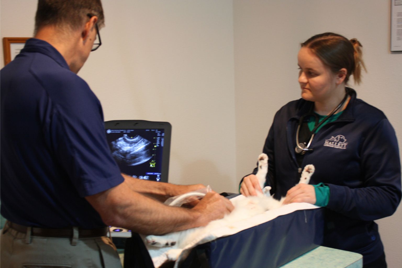 An ultrasound is the second most common type of diagnostic imaging tool veterinarians use to diagnose a cat's medical condition. Ultrasounds use soundwaves to examine and image internal tissues in real-time. An ultrasound allows a veterinarian to see into a cat's body (most commonly the abdomen), allowing for easy viewing of organs from different angles that are not easily achieved through x-rays. Ultrasound is also totally painless, but some cats can benefit from sedation to reduce anxiety and stress with positioning.
An ultrasound procedure usually proceeds as follows:
- The cat is placed in a padded trough/bed and is typically laying on it's back and held by a technician
- Sometimes the abdomen region needs to be shaved to obtain good images
- The veterinarian gently presses a small ultrasound probe against the cat's body that emits digital sound waves
MRI and CT scans
Magnetic resonance imaging, or MRI, is the newest form of diagnostic imaging being used for both human and veterinary medicine. MRI equipment generates a very powerful magnetic field, resulting in detailed anatomic images of whatever part of a cat's body is being scanned. No x-rays are involved, and an MRI is considered extremely safe. Cats must be anesthetized for this procedure because they cannot be restrained by humans and must remain still during the procedure
CT scans for cats, also known as "cat scans," are computer enhanced x-ray procedures most often used to evaluate complex parts of the body, such as the head, chest, some joints, and various internal organs. CT scans show different levels of tissue density and produce more detailed images than x-rays. Cats must be anesthetized for this procedure as well because they cannot be restrained by humans and must remain still during the procedure
Both CT and MRI are imaging modalities that many of the specialty practices in the area can perform on a referral basis. If one of our veterinarians is recommending one of these we will be able to get your cat referred, as we do not have the equipment to preform those at our hospital.
Veterinary diagnostic imaging offers an array of incredibly useful tools within a veterinarian's toolkit. Sometimes a diagnostic imaging session can lead to the need for further diagnostics.
Contact Us For Feline Radiographs or Ultrasound Today
If you are concerned that your cat might be injured or experiencing internal problems please schedule an appointment with one of our veterinarians today.